Range of Valley Fever Cases:
- Not apparent - 60%
- Mild to Moderate - 30%
- Complications - 5% to 10%
- Fatal - less than 1%
The usual course of disease in otherwise healthy people is complete recovery within six months.
In most cases, the body's immune response is effective and no specific course of treatment is necessary.
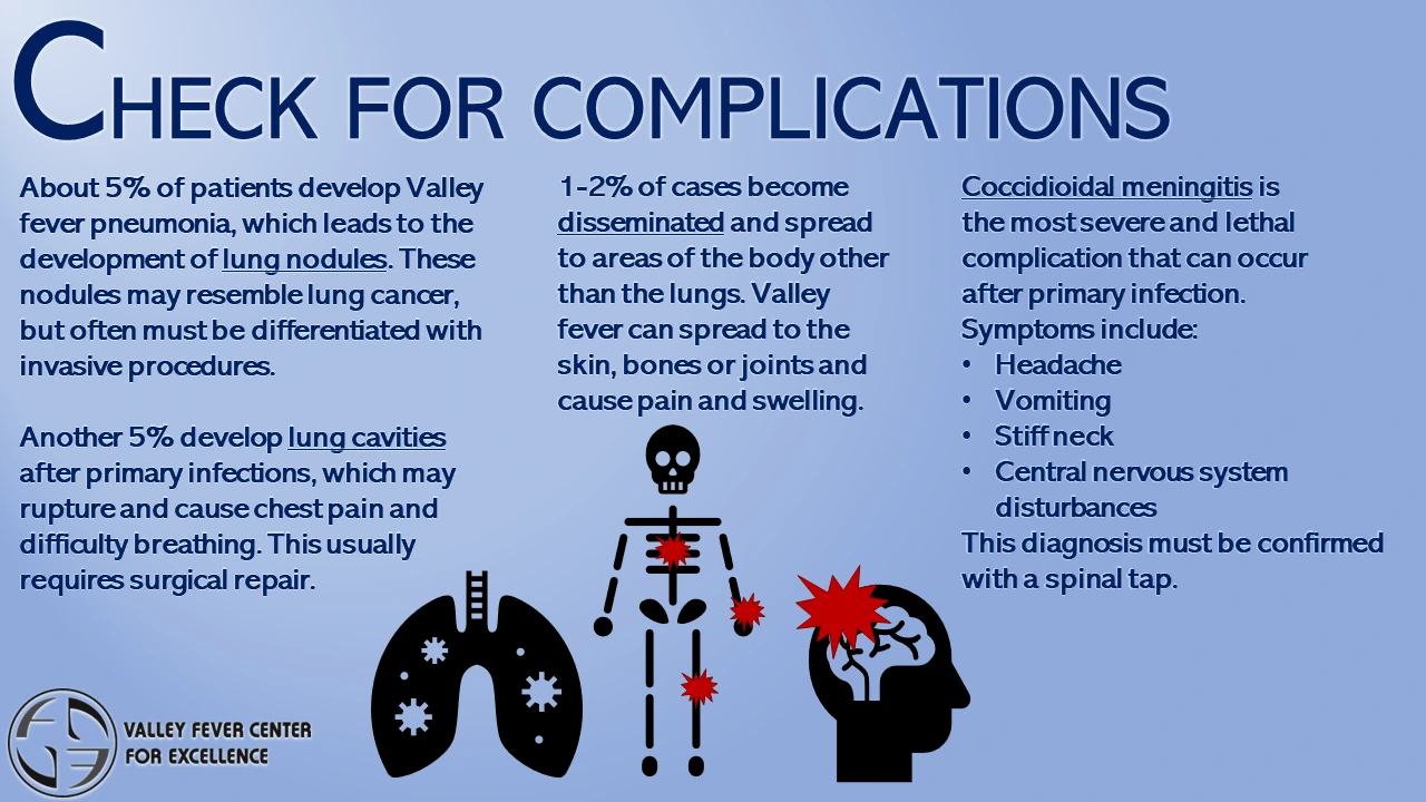
About five percent of cases of Valley Fever pneumonia (infection of the lungs) result in the development of nodules in the lung.
Nodules are small residual patches of infection that generally appear as solitary lesions, typically 1 - 1.5 inches in diameter, and often produce no symptoms. On a chest x-ray, these nodules resemble lung cancer. Unfortunately, it is usually not possible to make a definite diagnosis without removing a part or the entire nodule by bronchoscopy, needle-aspiration or surgery.
Another five percent of patients develop lung cavities after their initial infection with Valley Fever.
These cavities occur most often in older adults, usually without symptoms, and about 50% of them disappear within two years. Occasionally, these cavities rupture, causing chest pain and difficulty breathing, and require surgical repair.
Of those patients who seek medical attention, one to two percent develop disease that has spread (disseminated) to other parts of the body.
The most common site of dissemination is the skin. Biopsies of skin lesions may reveal Coccidioides when grown in culture.
Bones and joints (especially the knees, vertebrae, and wrists) are frequent sites of dissemination.
The changes in bones and joints due to Valley Fever can be seen on x-rays and in CT-scans of the affected body part.
Meningitis is the most serious and lethal complication of disseminated disease.
Symptoms include headache, vomiting, stiff neck, and other central nervous system disturbances. A spinal tap is required for a definite diagnosis of meningitis.
CLINICAL IMAGES OF COMPLICATIONS

